02 Apr, 2019
KONICA's MX1
Good things come in small packages!
Konica Minolta’s MX1
The number of ultrasound machines that are being launched leaves one in a state of confusion, just like the avalanche of SUV’s in 2019!
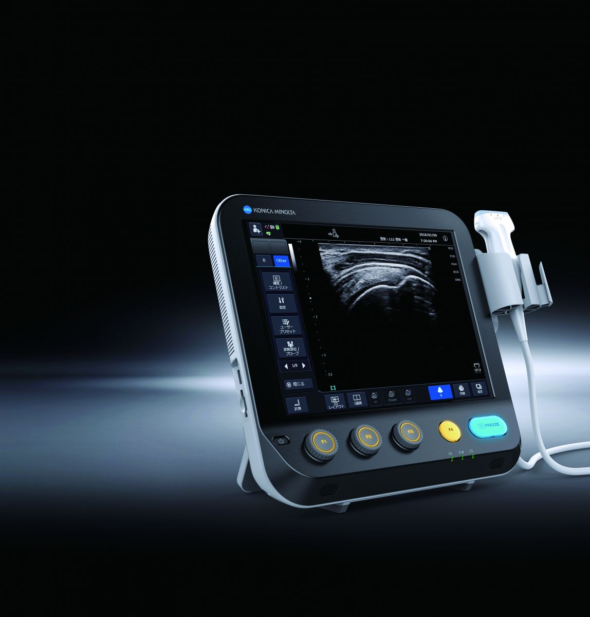
But amongst the multiple machines, is one that comes as a breath of fresh air. Compact, easy on the pocket, and with a surprisingly amazing clarity for a machine in the portable class, is Konica’s new SONIMAGE MX1. The MX1 is a new Point-of-Care system that inherits a DNA of innovation from the higher-end model SONIMAGE HS1.
EXTERIOR
It’s a pocket sized dynamite, weighing in at a light 4.5 kg, and has a nice wide 12.1 inch touchscreen. At the bottom of the machine are 5 keys, that are used most frequently, thus allowing for improved efficacy and thoroughput. It comes with 4 probes, that include a convex, 2 linear options, and one microconvex. It has a Cradle, a mounting platform for MX1 that allows to hand-carry the system without insertion and removal of AC power/USB cable. It can be mounted on the cart with VESA and enables to charge the battery when you put the system back.
WORKFLOW
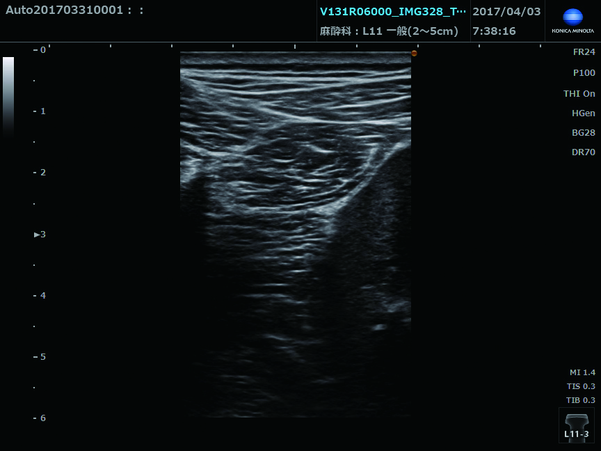
The machine looks light, but has a tremendous work rate and efficacy that is sure to make the competition sit up and look. The machine can run on the battery pack for 2 hours easily, and one can always put the machine back on the cart for charging if its running low. This way, one can move the machine across intensive care and different work areas without switching off and on. The musculoskeletal applications with the L11-3 probe are wonderful, providing exceptional image quality from deep area to superficial and visualization of biopsy needle as well. Using a special Triad Tissue Harmonics, the machine is valuable for superficial structures as well.
Another important feature is the Multi Parameter Adapter (MPA) that enables to change multiple image parameters like frequency change and turning trapezoid on in conjunction with depth change. This feature reduces unnecessary keystrokes to optimize the image and hence increases throughput. Multi image Display is common in many machines, but this machine allows one to compare stored images with real time images side by side, so one can check resolution of lesions and response to treatment on a real-time basis.
SUMMARY
This machine surprises all with his high quality images, especially the L11-3 probe for superficial, musculoskeletal and Doppler applications, as well as DNA and biopsy needle visualization. It is likely to carve a niche for itself in busy intensive care units, point of care situations, as well as a second line machine in Radiology clinics with a busy workload.