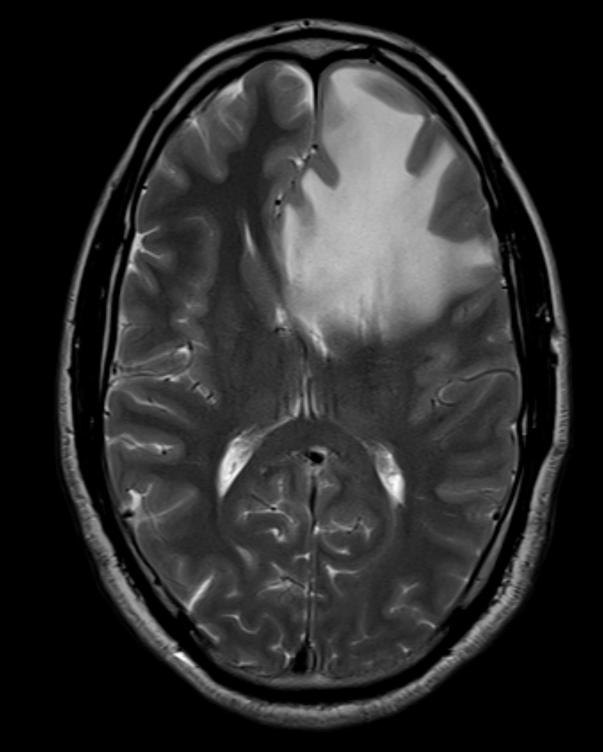
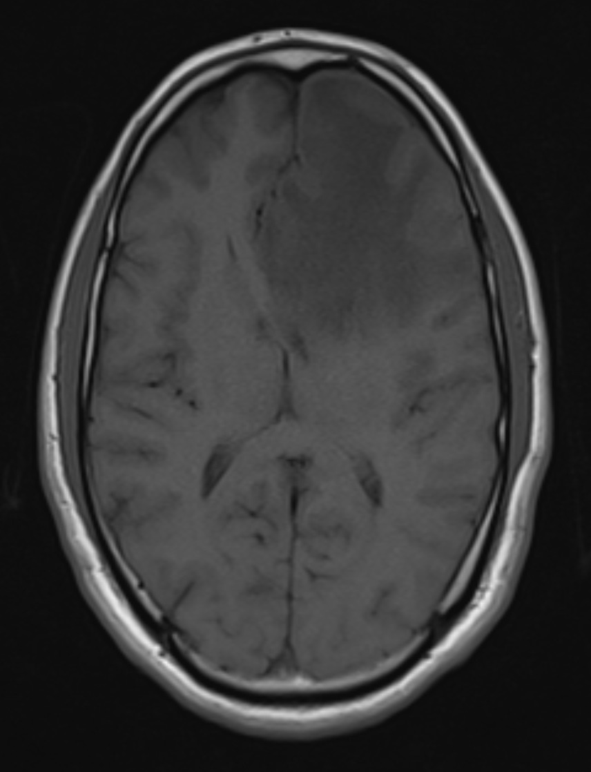
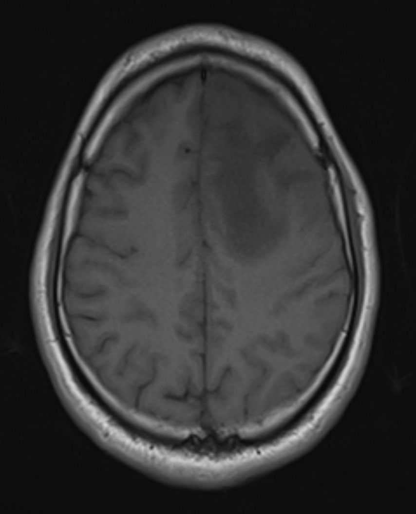
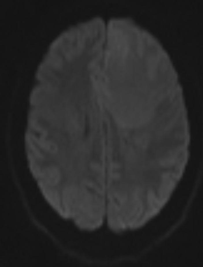
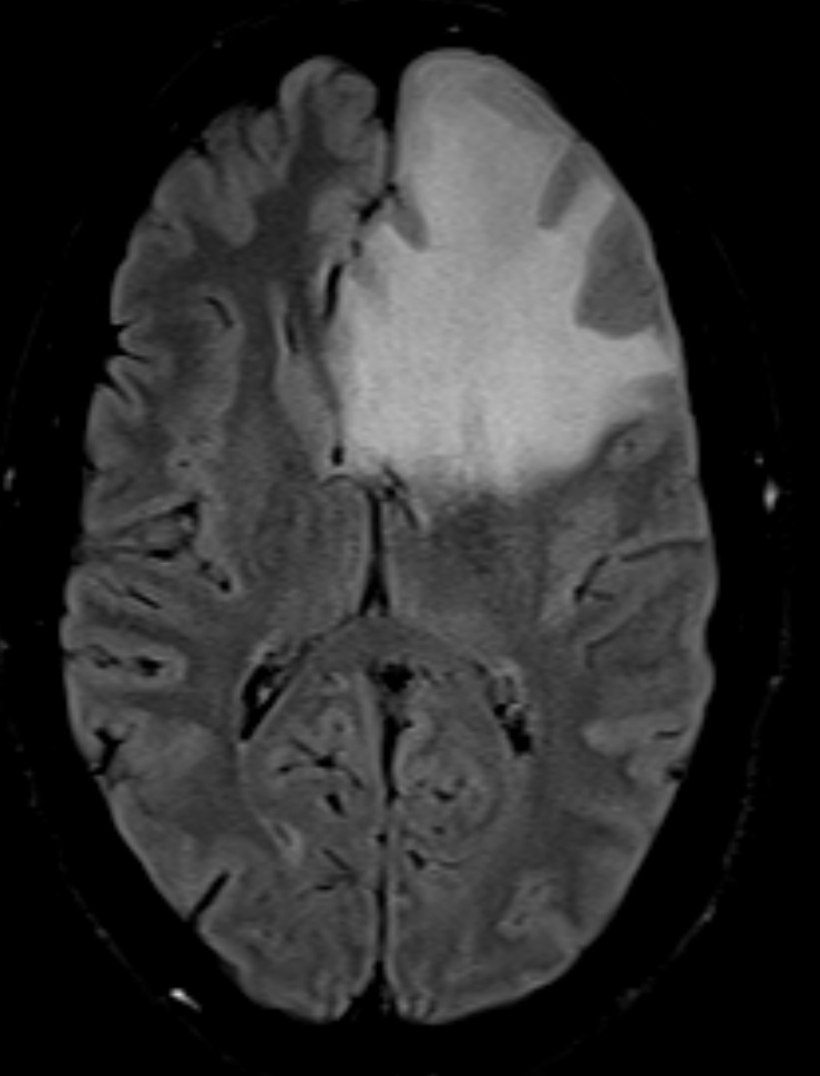
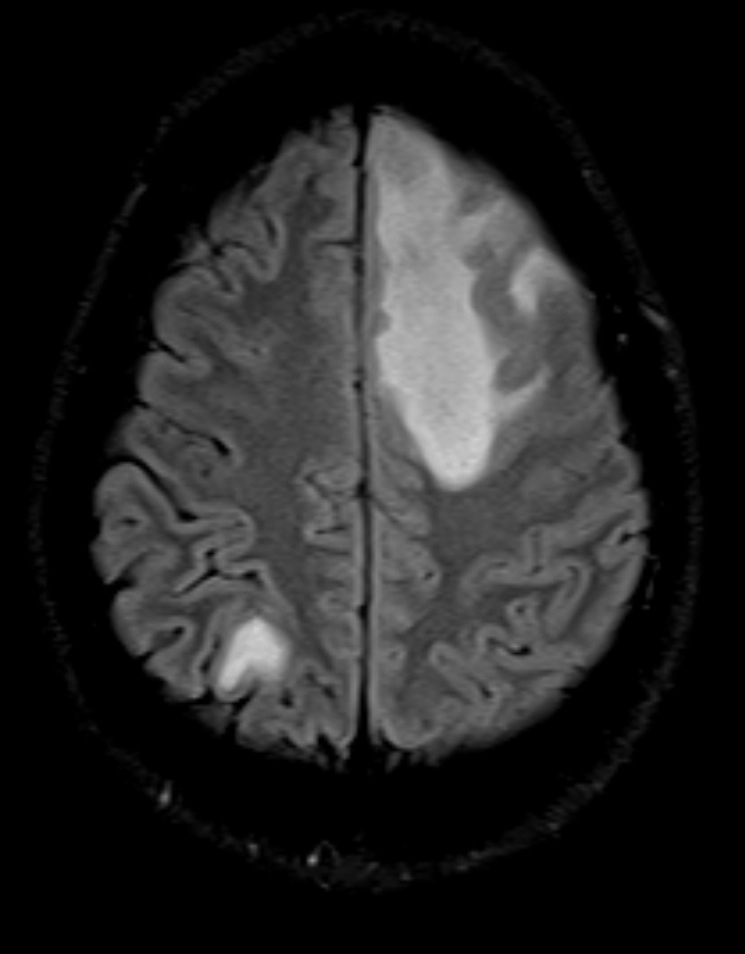
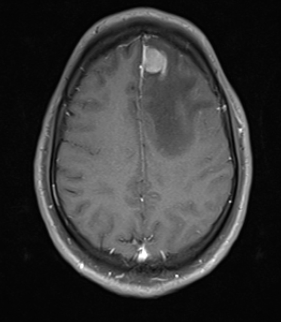
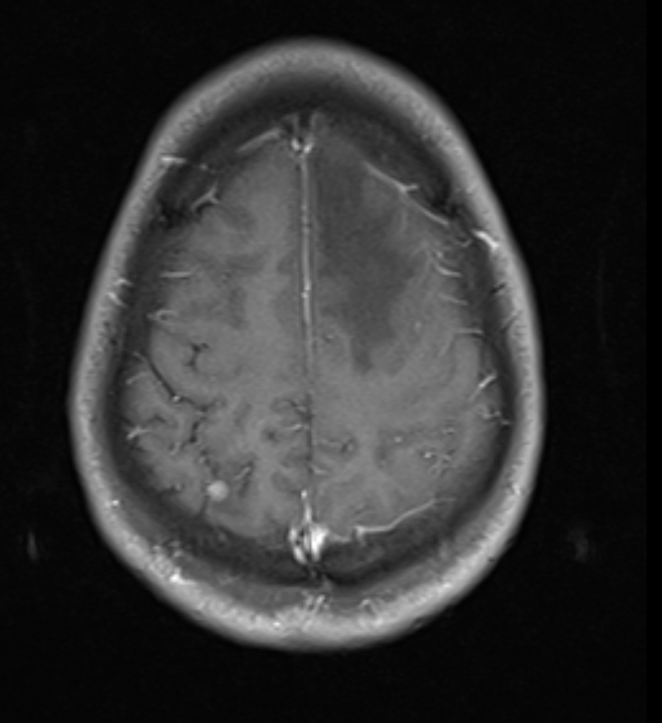
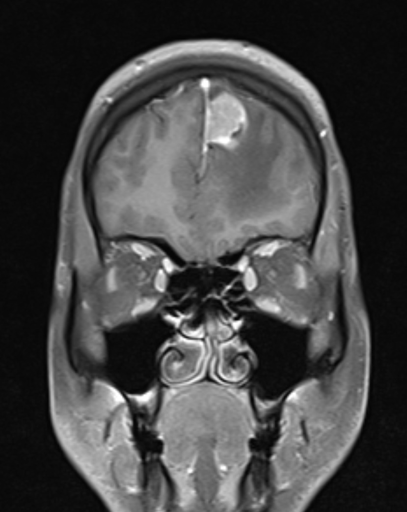
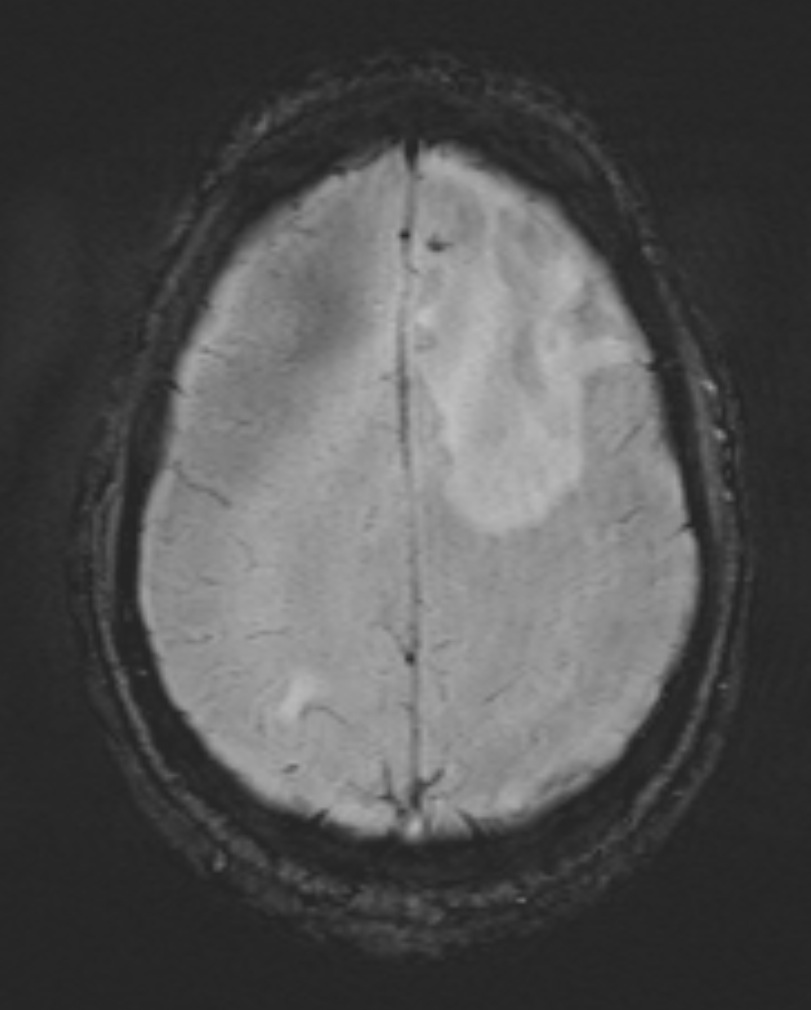
STUDY reveals homgenously enhancing lesions in the left para falcine frontal region and right parietal region with surrounding edema and mass effect. The left para falcine lesion shows a "dural tail' like apperance, that is though usually seen in meningiomas, but can be seen in any dural based tumors, especially mets. A speck of hemorrhage is also noted in this lesion on SWI sequences.
The lesions are not ring enhancing or conglomerate, hence rules out tuberculomas